Signal Characteristics, Noise, and Low SNR
Overview
A central fact about EEG is that the useful neural signal is weak relative to many nuisance sources. This is the low signal-to-noise ratio (SNR) problem. Any serious EEG pipeline, whether classical or deep-learning-based, must be designed with this issue in mind.
Why EEG Has Low SNR
The measured scalp potential is the result of several attenuating and mixing processes:
- neural activity is spatially aggregated,
- the skull acts as a low-conductivity barrier,
- scalp and environmental sources contaminate recordings,
- reference choices can reshape apparent signal structure.
As a result, emotionally relevant brain activity may be subtle compared with noise and artifacts.
Common Noise and Artifact Sources
Physiological Artifacts
- EOG: eye blinks and eye movements
- EMG: facial and neck muscle activity
- ECG leakage: cardiac contamination in some channels
- Motion artifacts: electrode shifts and cable movement
Environmental Artifacts
- line noise at 50/60 Hz,
- poor shielding or grounding,
- nearby electronics,
- impedance instability.
Experimental and Behavioral Confounds
- subject fatigue,
- attention fluctuation,
- posture changes,
- task noncompliance,
- emotional self-report inconsistency.
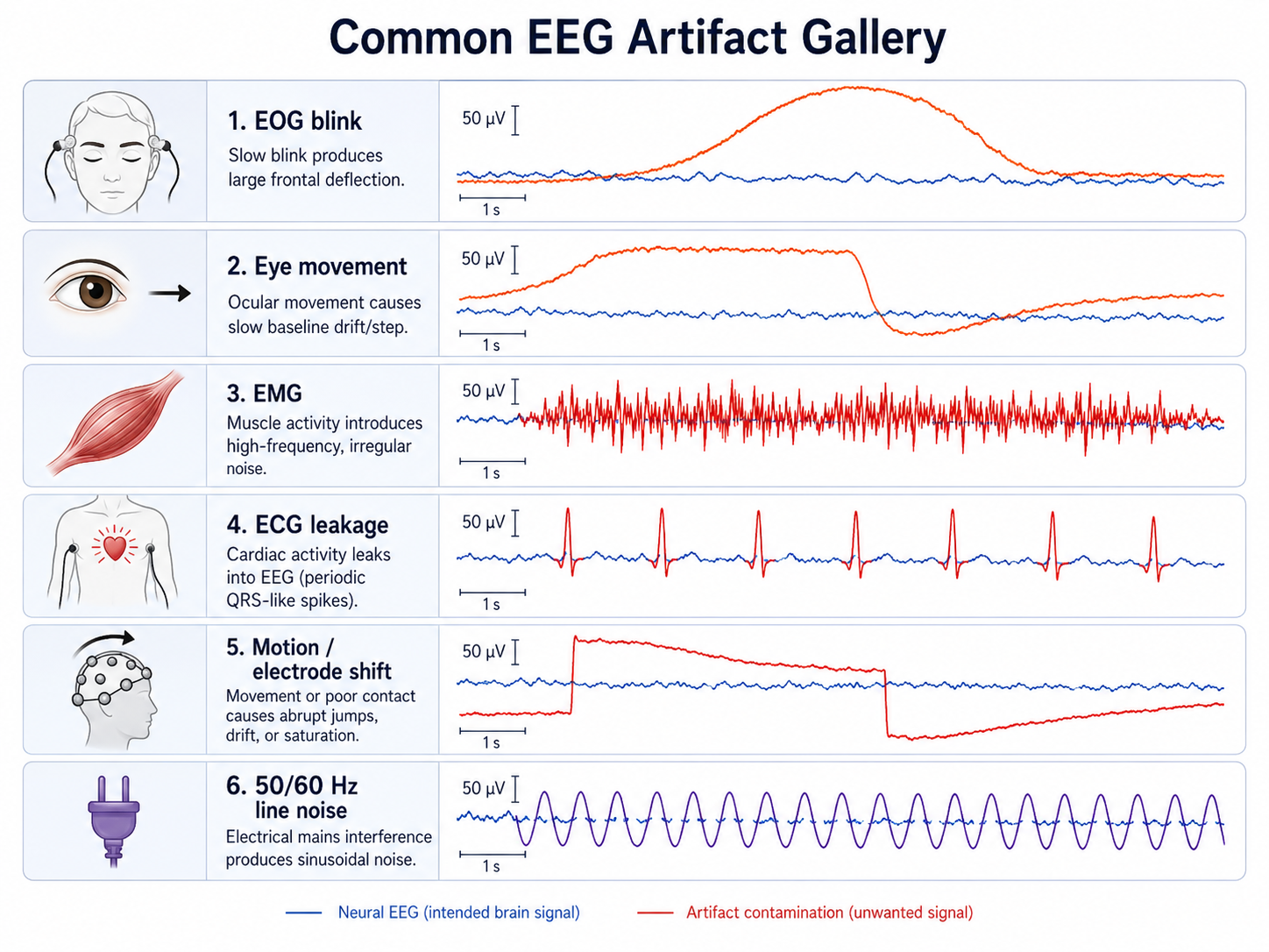
Figure 1.7: Artifact gallery. Aligned EEG traces illustrate common contamination sources, including EOG blinks and eye movements, EMG, ECG leakage, electrode or cable motion, and 50/60 Hz line noise. The traces are paired with source labels so physiological, environmental, and movement artifacts can be distinguished from neural signal.
Signal-to-Noise Ratio
SNR is often described conceptually as
Low SNR means that the neural activity of interest contributes only a small fraction of the observed variance.
In affective computing, this is especially problematic because emotional effects are often subtler than motor or visual evoked responses.
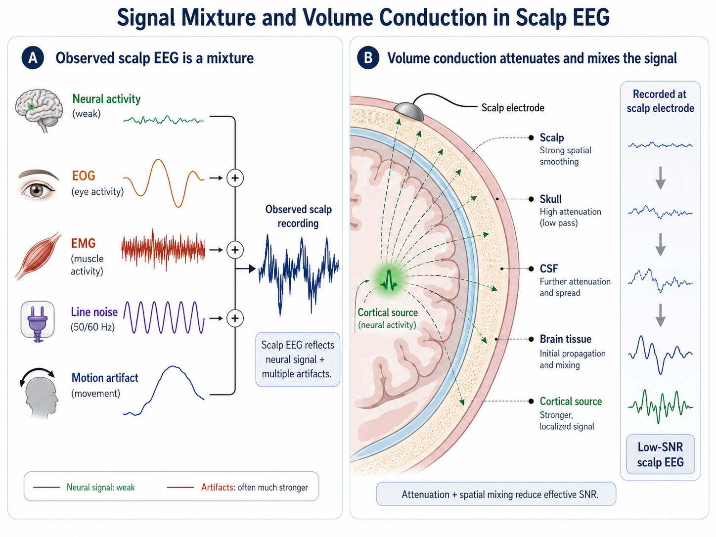
Figure 1.8: Signal mixture and volume conduction. A weak neural component is combined with EOG, EMG, line noise, and motion artifacts to form the observed scalp recording. A second panel shows attenuation and spatial mixing as the signal passes through brain tissue, cerebrospinal fluid, skull, and scalp, yielding a low-SNR waveform at the electrode.
EEG Frequency Bands
Although exact boundaries vary across studies, EEG is commonly analyzed in bands such as:
- Delta: roughly 0.5–4 Hz
- Theta: roughly 4–8 Hz
- Alpha: roughly 8–13 Hz
- Beta: roughly 13–30 Hz
- Gamma: above 30 Hz
These bands are useful abstractions, but low SNR means that estimated band power can be strongly affected by artifacts and preprocessing decisions.
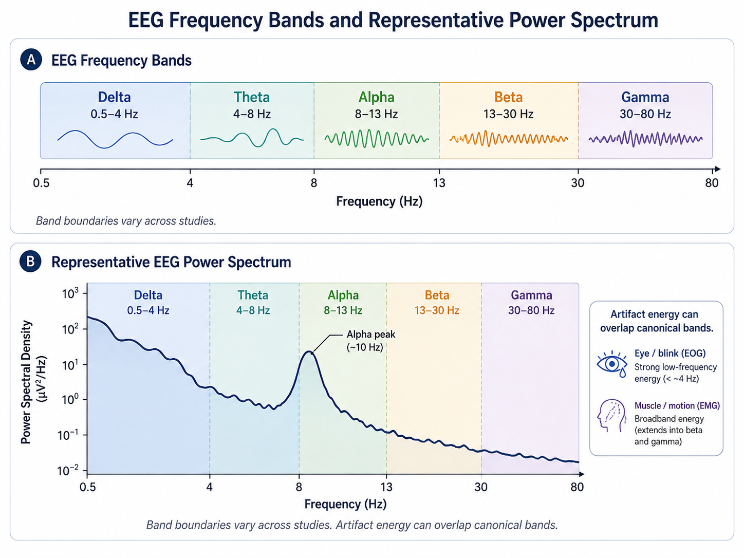
Figure 1.9: EEG frequency bands and power spectrum. The horizontal axis spans the typical EEG range from delta (about 0.5–4 Hz) through theta, alpha, beta, and gamma. Each band is marked along the axis and illustrated with a representative power spectrum. Exact boundaries vary across studies, and artifact energy can overlap the bands, so interpretation should be paired with careful preprocessing.
Why Low SNR Matters for Modeling
Low SNR affects downstream learning in several ways:
- models may overfit artifact patterns,
- useful features may be swamped by nuisance variation,
- deeper models may amplify noise if data are limited,
- subject transfer becomes harder because nuisance structure differs across people.
Thus, EEG modeling is never only a modeling problem. It is also a signal quality problem.
Strategies to Improve Effective SNR
During Acquisition
- improve electrode contact quality,
- reduce motion and environmental interference,
- use appropriate referencing,
- monitor impedance,
- design tasks that reduce unnecessary movement.
During Preprocessing
- bandpass filtering,
- notch filtering,
- artifact rejection,
- ICA or related decomposition methods,
- rereferencing,
- segmentation and baseline correction.
During Modeling
- robust feature extraction,
- channel selection,
- regularization,
- subject-aware validation,
- augmentation that respects signal structure.
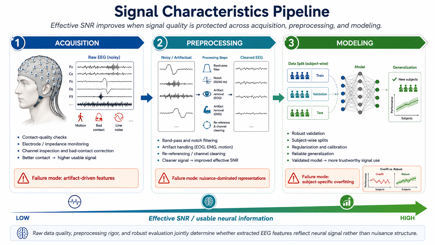
Figure 1.10: Three-stage SNR pipeline. Acquisition, preprocessing, and modeling each offer opportunities to improve effective signal-to-noise ratio. Contact-quality checks and clean acquisition reduce physiological and environmental artifacts at the start. Filtering, artifact rejection, ICA, rereferencing, and baseline correction further separate signal from noise. Robust feature extraction, regularization, and subject-aware validation keep downstream models from overfitting nuisance patterns or subject-specific artifacts.
Event-Related vs. Ongoing EEG
SNR can be improved by averaging when responses are time-locked and repeatable, as in classical event-related potential studies. But affective computing often uses more continuous and naturalistic paradigms, where averaging is less straightforward. This is one reason why low SNR remains a central challenge for emotion-related EEG.
Summary
EEG has inherently low SNR because weak neural signals are mixed with substantial physiological, environmental, and experimental noise. Understanding this constraint is essential for later decisions about preprocessing, feature representation, and deep model design.
Next: Electrodes, Montages, Wearables, and Source Estimation
References
- Urigüen, J. A., and Garcia-Zapirain, B. (2015). EEG artifact removal—State-of-the-art and guidelines. Journal of Neural Engineering, 12(3), 031001.
- Muthukumaraswamy, S. D. (2013). High-frequency brain activity and muscle artifacts in MEG/EEG: A review and recommendations. Frontiers in Human Neuroscience, 7, 138.
- Delorme, A., Sejnowski, T., and Makeig, S. (2007). Enhanced detection of artifacts in EEG data using higher-order statistics and independent component analysis. NeuroImage, 34(4), 1443–1449.
- Klimesch, W. (1999). EEG alpha and theta oscillations reflect cognitive and memory performance: A review and analysis. Brain Research Reviews, 29(2–3), 169–195.
- Luck, S. J. (2014). An Introduction to the Event-Related Potential Technique (2nd ed.). MIT Press.
- Cohen, M. X. (2014). Analyzing Neural Time Series Data: Theory and Practice. MIT Press.